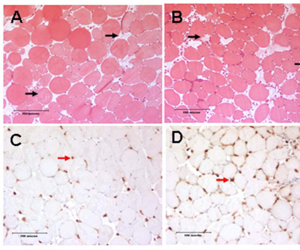
Figure 1. Histologic assessment of hind limb acute muscle fiber injury and neutrophil infiltration following acute IR.
Images A and B are mason trichrome stained TA muscle cross sections taken from DIO or ND mice respectively obtained 24 hours following IR. Black arrows indicate injured skeletal muscle fibers in each field. Images C and D are Ly6B positive neutrophils (red arrow) in the TA muscle, taken from DIO and ND mice respectively. There was no difference in the average number acute of injured fiber between the two groups; however we identified markedly more infiltrating neutrophil granulocytes in the DIO muscles 24 hours after IR.

