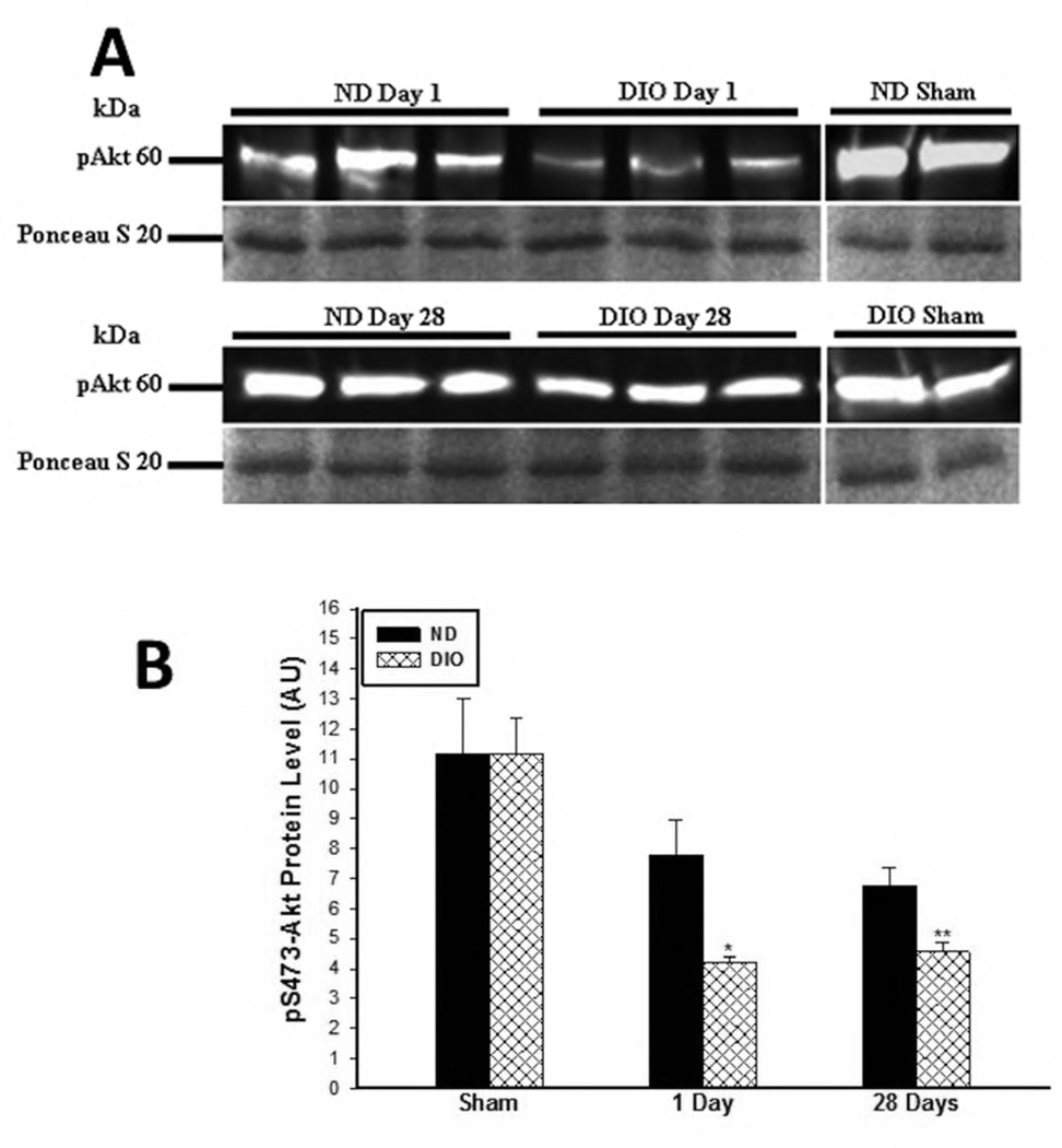
Figure 5. A. Representative western blot image for phosphorylated-S473-Akt in soluble skeletal muscle protein samples obtained at 1 and 28 days following IR. B. Assessment of Akt pathway activity at 1 and 28 days following IR.
There was no difference in phosphorylated Serine 473-Akt under sham condition. However by 1 and 28 days after IR there was significantly lower pS473 expression in the DIO mice or 28 days following IR compared to ND (*,** p<0.01, n=6–7).

