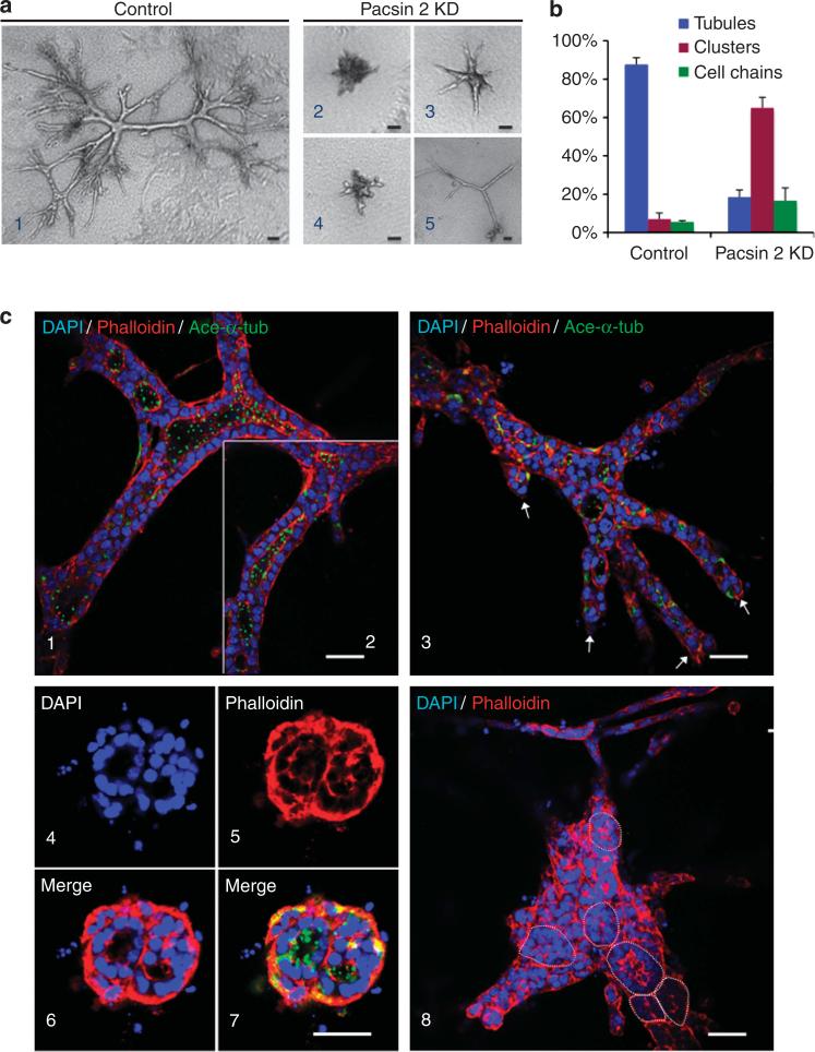
Figure 5. Pacsin 2 depletion leads to defects in tubulogenesis in three-dimensional (3D) collagen gels.
(a) Phase-contrast images for (1) control and (2–5) Pacsin 2 knockdown murine inner medullary collecting duct 3 (mIMCD3) cells cultured for 12 days in 3D type I collagen gels. (b) Quantification of structures formed in 3D tubulogenesis assays. The structures formed in 3D collagen gels were classified into three categories (tubule, cell cluster, and cell chain). The percentage of each type of the structures was calculated by dividing by total number of structures formed. Cell structures were counted from four individual experiments. More than 160 structures were counted for each cell type. Error bars represent±s.d. (c) Confocal images of cells cultured in a stained with phalloidin and acetylated α-tubulin (ace-α-tub; green). DAPI, 4’,6-diamidino-2-phenylindole. (1–2) Control mIMCD3 cells formed large elaborately branched tubular structures with well-established lumen lined with a single layer of epithelial cells with apical primary cilia protruding toward the lumen. The lumen for the tubule in (1) and (2) are connected, shown by two confocal optical sections that are 2 μm apart in the focal plane. (3–8) Pacsin 2 knockdown cells have defects in tubule formation. They tend to form tubules with blunt ends (3, arrows point to the blunt ends), or cell clusters with isolated lumens or multi-lumens (4–8). (4–7) Typical multilumen structures seen in Pacsin 2 knockdown mIMCD3 cells. (8) A large structure with at least six isolated lumens. Circle outlines the lumen. Bar = 50 μm.

