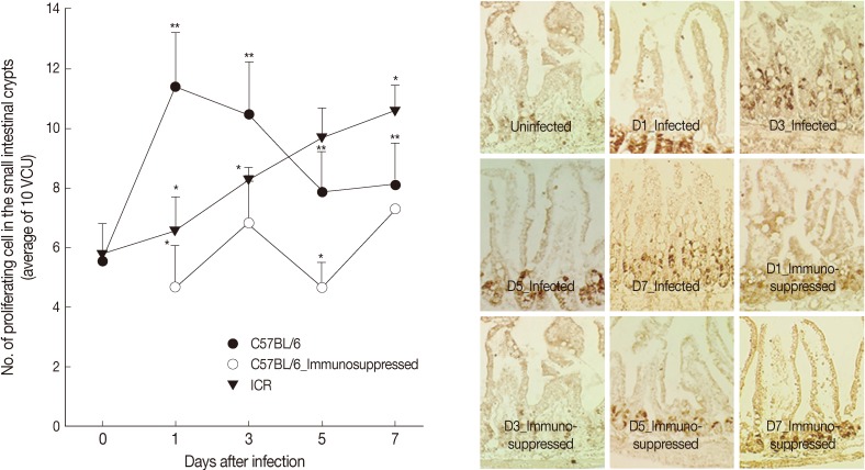
Fig. 3.
Intestinal epithelial cell turnover of C57BL/6 and ICR mice. (A) Intestinal epithelial cell turnover was measured in C57BL/6 (immunocompetent and immunosuppressed) and ICR mice infected with 200 G. seoi metacercariae. The number of newly proliferating intestinal epithelial cells in the crypt was counted at 10 villus-crypt unit and averaged. Statistically significant differences (P<0.05) compared with C57BL/6 mice were expressed as an asterisk (*). In immunocompetent C57BL/6 mice, dark brown-stained epithelial cell counts at day 1 (**), day 3 (**), day 5 (**), and day 7 (**) were significantly higher (P<0.05) than the figure of uninfected C57BL/6 mice (day 0). (B) Changes of epithelial cell turnover in immunocompetent and immunosuppressed C57BL/6 mice in the middle part of the jejunum infected with 200 G. seoi metacercariae at days 0 (uninfected), 1, 3, 5, and 7 PI. The nuclei of proliferating epithelial cells are stained dark brown by bromo-2'-deoxyuridine (BrdU) immunohistochemical staining.

