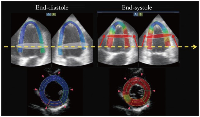
Fig. 8.

Out of plane phenomenon. Upper panels are 4 chamber and 2 chamber views at end-diastole and systole in a healthy subject. The color shows degree of longitudinal displacements during systole. White discs at end-diastole are moved to red discs level at end-systole, which distance is about 15 mm (red arrows). Lower panels show the 2-dimensional speckle tracking echocardiography (2D-STE) images at yellow dash arrow level. Since the white disc level is moved to apex at end-systole, the 2D-STE image at end-systole is changed to another plane of a basal level at end-diastole. Such changing of the plane of interest through a cardiac cycle is called as out of plane phenomenon.
