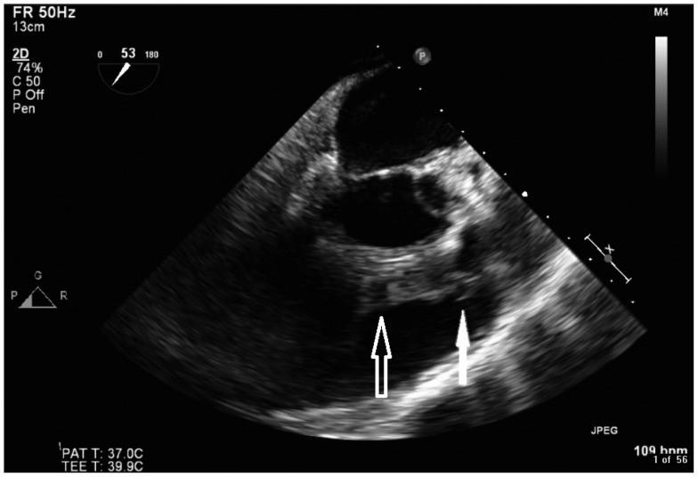
Fig. 3.
Transoesophageal echocardiogram. Mid-oesophageal right ventricular outflow view showing large sized vegetations attached to the right ventricular outflow tract (RVOT) side of the pulmonic valve (solid arrow). An abscess within the posterior wall of the RVOT, note the echo free spaces within (open arrow).

