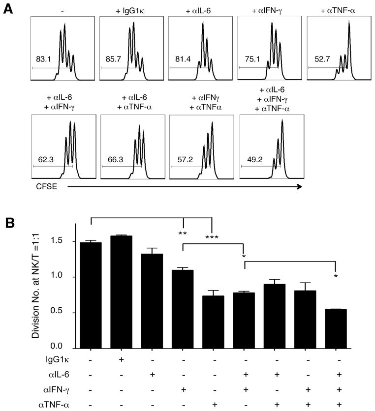
Figure 6. Neutralizing IFN-γ, TNF-α, and IL-6 in NK-T cell co-culture ameliorated CD4+ T cell proliferation.
NK and autologous CD4+ T cells were isolated from an AA haplotype licensed individual, stimulated with anti-CD3 and anti-CD28, and co-cultured in 2 ng mL−1 (26 I.U) IL-2 for 3 days. (A) Histograms of CD4+ T cell CFSE dilution without or with the indicated neutralizing antibodies. The number in each histogram indicates the percentage of cells proliferated (gated on CD4+CFSE+ cells). (B) Bar plot of CD4+ T cells division number at NK/T = 1:1 from the AA haplotype healthy individual. (Mean ± SEM, n = 2 to 6, two-tailed student t test, * p < 0.05; ** p < 0.005; *** p < 0.0005). More than three experiments were performed.

