
Figure 5. Long-term Smoothened inhibition yields poorly differentiated pancreatic tumors with increased proliferation and vascularity.
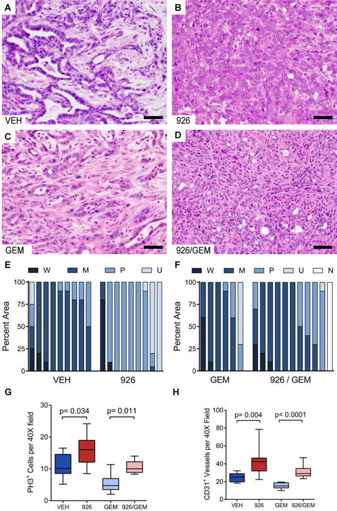
(A–D) Representative histology (hematoxylin and eosin stain) of KPC tumors arising after long-term treatment with vehicle (A), IPI-926 (B), gemcitabine + vehicle (C), or gemcitabine + IPI-926 (D).
(E and F) Quantification of the differentiation state for each cohort. The fraction of each tumor that was observed to be well-differentiated (W), moderately differentiated (M), poorly-differentiated (P), or undifferentiated (U), was scored in a blinded manner, and compared between the treatments. No tumor could be located in one IPI-926 + gemcitabine treated mouse (N).
(G) Quantification of phospho-histone H3+ cells per 40X field in each treatment group (p=0.034 for monotherapy, p=0.011 for combination). Data are presented as standard box and whisker plots.
(H) Quantification of CD31+ vessel structures by IHC in each treatment cohort (p=0.004 for monotherapy, p<0.0001 for combination). Data are presented as standard box and whisker plots.
Two-tailed Mann-Whitney U was used for all unpaired tests. Scale bars = 50 μm.
See also Figure S4.