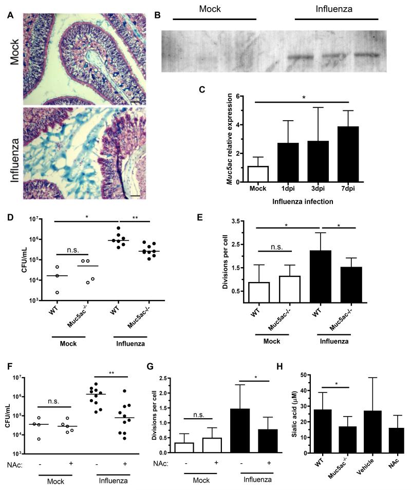
Figure 4. Sialylated airway mucins are required for influenza-induced pneumococcal growth.
(A) Mouse heads were obtained after 7 days of mock or influenza infection, fixed, decalcified and sectioned. URT sections were stained with alcian blue and nuclear fast red. Scale bars, 50 μm. (B) Nasal lavages were obtained from mice infected with influenza or PBS for 7 days, then analyzed by Western blot for the presence of Muc5ac. (C) Nasal lavages with RLT RNA lysis buffer were obtained from mice influenza or mock-infected for 7 days. qRT-PCR was performed, and relative expression of Muc5ac measured. (D) Mice of indicated genotype were mock or influenza infected for 7 days, followed by inoculation with CFSE-labeled pneumococci for 8 hrs. Nasal lavages were obtained and plated for quantitative culture. (E) Lavages were also fixed, stained for pneumococcal capsule, and flow cytometry performed to measure bacterial replication (division index). (F-G) Wildtype mice were infected with influenza or mock, followed by daily treatment for 7 days with vehicle (PBS) or 0.5 M N-acetylcysteine (NAc). CFSE-labeled pneumococci were inoculated for 8 hrs, then nasal lavages obtained and used for quantitative culture (F) and flow cytometric analysis of cell division (G). (H) Total sialic acid content was measured by thiobarbituric acid assay on samples from D-G. Data are represented as mean +/− SD. Horizontal lines indicate median values. * = p < 0.05, ** = p < 0.01. Experiments were performed at least twice, with 3-13 mice per group.

