
Figure 2.
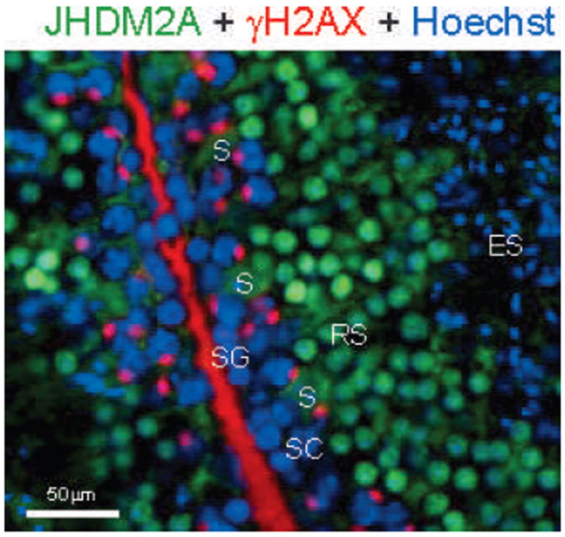
Immunohistochemical anaylsis of JmjC domain–containing histone demethylase 2A (JHDM2A) in mouse testis. JHDM2A (green) is highly expressed in round spermatids (RS) and not quite merged with gamma histone 2A variant X (γH2AX)-positive (red) spermatogonia (SG) and spermatocytes (SC). JHDM2A-positive signals disappear in elongated spermatids (ES). Cytoplasmic staining of JHDM2A is observed in Sertoli cells (S). Hoechst (DNA) staining is shown as blue.