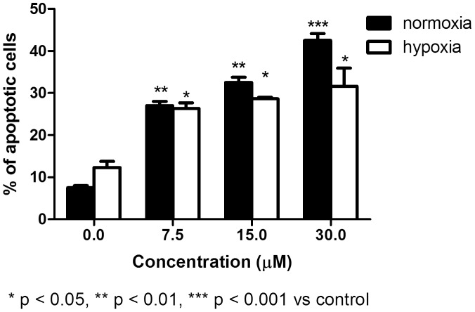
Figure 4. Induction of apoptosis by SR in normoxic and hypoxic conditions on REH cells.
Cells were treated with 0 to 30 µM SR for 24 h. The % of apoptotic cells recorded in the untreated cultures was subtracted from that observed in cultures treated with SR. Data are presented as mean ± SEM of at least four different experiments.

