Figure 1.

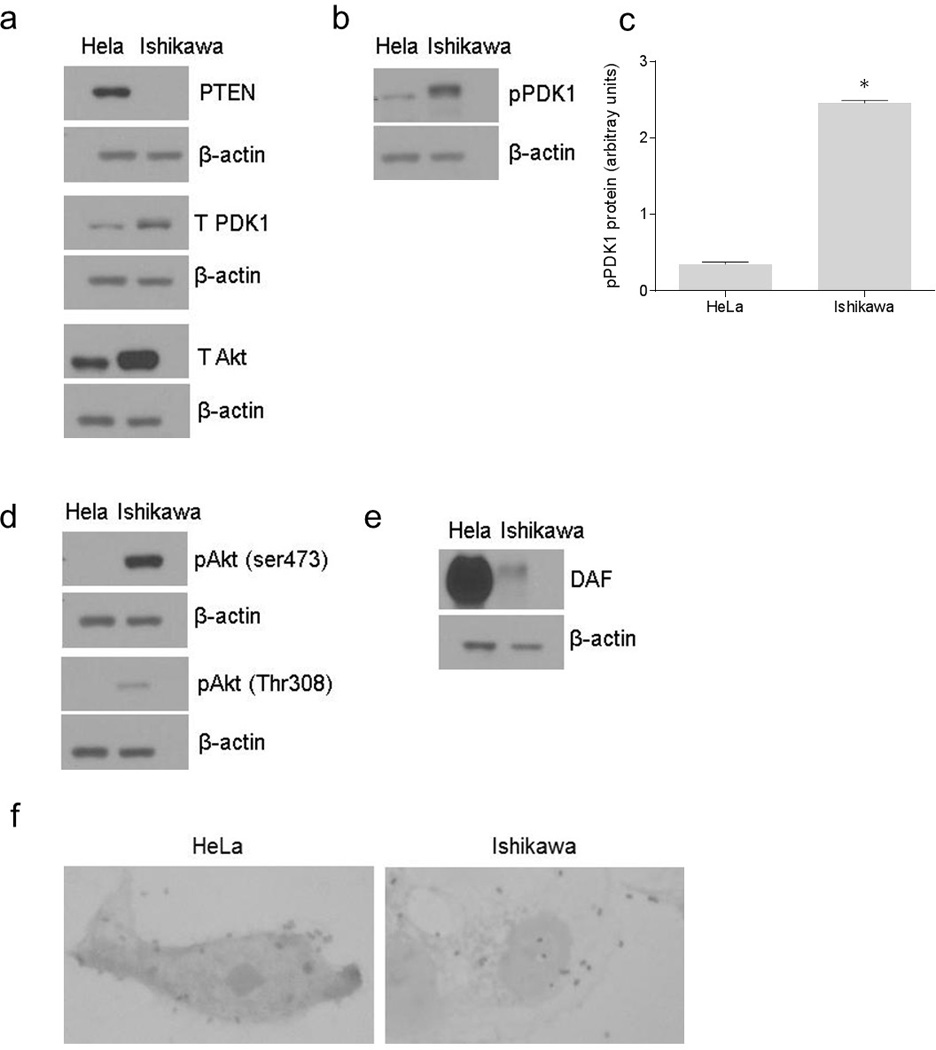
The association between constitutive activity of PI3K/Akt pathway and adhesion of Dr+ E. coli to epithelial cells. The expression of PI3K/Akt pathway proteins in HeLa and Ishikawa cell grown as monolayers for 24 hours was examined by western blot. The E. coli K-12 strain carrying the plasmid pBJN406, encoding the production of Dr fimbriae was allowed to bind to HeLa and Ishikawa cells for three hours and then the number of bacteria adhered to individual cells was examined by gram staining. (a) Western blot for PTEN, total PDK1 and total Akt proteins in HeLa and Ishikawa cells. (b) Western blot for pPDK1 protein in HeLa and Ishikawa cells. (c) Densitometric analysis of pPDK1 protein levels. * p < 0.0001 versus HeLa. (d) Western blot for activated forms of Akt, pAkt(ser473) and pAkt(Thr308). (e) Western blot for DAF. (f) Gram staining to visualize E. coli K-12 strain carrying plasmid pBJN406 adhered to HeLa and Ishikawa cells. Data are representative images or expressed as mean values ± SEM of each group from three separate experiments.
