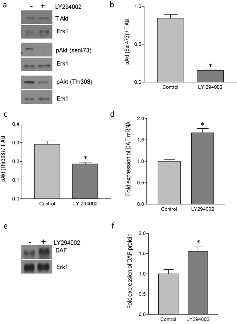
Figure 2.

Up-regulation of DAF by pharmacological inhibitor of PI3K/Akt pathway. The monolayer of Ishikawa cells was treated with LY 294002 (50 µM) dissolved in DMSO or DMSO alone (control) for 24 hours. (a) Western blot for total Akt and its activated forms, pAkt(ser473) and pAkt(Thr308). (b) Density analysis of pAkt(ser473) western blot expressed as ratio between total Akt and pAkt(ser473). * p < 0.0001 versus control. (c) Density analysis of pAkt(Thr308) western blot expressed as ratio between total Akt and pAkt(Thr308). * p < 0.01 versus control. (d) Fold increase in the expression of DAF mRNA as assessed by real time qPCR after treating the Ishikawa cells with the LY294002. * p < 0.001 versus control. (e) Western blot for DAF protein (f) Density analysis of DAF western blot expressed as fold increase in the expression after treating Ishikawa cells with LY294002. p < 0.03 versus control. Data are representative images or expressed as mean values ± SEM of each group from three separate experiments.
