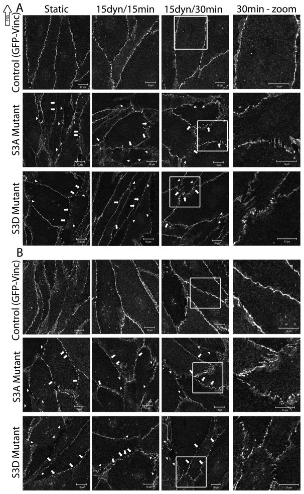
Figure 6.
The role of cofilin in FSS-induced barrier staining
BAECs were electroporated with 20μg/ml of either S3A or S3D cofilin constructs in combination with GFP-vinc as a fluorescent marker of transfection efficiency as described in the methods. BAECs expressing the mutant constructs were exposed to 15 dynes/cm2 FSS for 15 and 30mins and labeled using antibodies against VE-cadherin [A] or β-catenin [B]. The images shown are representative of 3 repeats. In both A and B, the top panel images are expressing GFP-vinc alone, the middle panel images are BAECs expressing S3A and GFP-Vinc, and the bottom panel images are BAECs expressing S3D and GFP-vinc. The three left columns are magnified 2X the original. The rightmost column is magnified 4X the original and highlights the boxed area in the column immediately to the left. Arrows point to small breaks in staining. Arrowheads point to large gaps in staining. Scale bars = 10μm.

