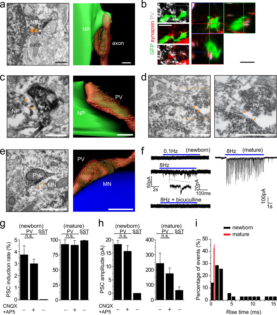
Figure 1.
PV+ interneurons form immature synaptic inputs onto proliferating newborn progeny in the adult dentate gyrus. (a) Sample immuno-EM image of a symmetrical synaptic contact (arrows) with a dark labelled newborn progeny (NP; left; Scale bar: 0.2 µm) and the serial reconstruction (right; Scale bar: 0.8 µm). (b) Sample confocal images of immunostaining for PV, synapsin I, and GFP (left) with orthogonal views (right). Scale bars: 20 µm. See Supplementary Movie 1 for 3D reconstruction and Supplementary Fig. 3 for another example for a proliferating newborn progeny. (c-e) Sample images and serial reconstructions of symmetrical synaptic contacts (arrowheads) between vesicle-filled PV+ axon terminals and newborn progeny (NP; c, d) or unlabelled mature neurons (MN; e). Please see Supplementary Fig. 4 for correlative light and EM analyses for the identification of labelled PV+ axonal terminals, labelled newborn progeny and unlabeled mature granule cell shown in (c, e). Scale bars: 1 µm. (f) Sample traces of whole-cell voltage-clamp recording of a RFP+ new progeny and a mature granule cell in the same preparation upon light stimulation (472 nm; 5 ms) of ChR2-expressing PV+ neurons. (g-i) Summaries of induction rate (g), mean amplitude (h), and distribution of rise times (i) of evoked PSC recorded from RFP+ newborn progeny at 4 dpi and from mature granule neurons upon 8 Hz light stimulation of ChR2-expressing PV+ or SST+ neurons. Values represent mean ± s.e.m. (n = 3–8 cells; n.s.: P > 0.1; Student’s t-test).

