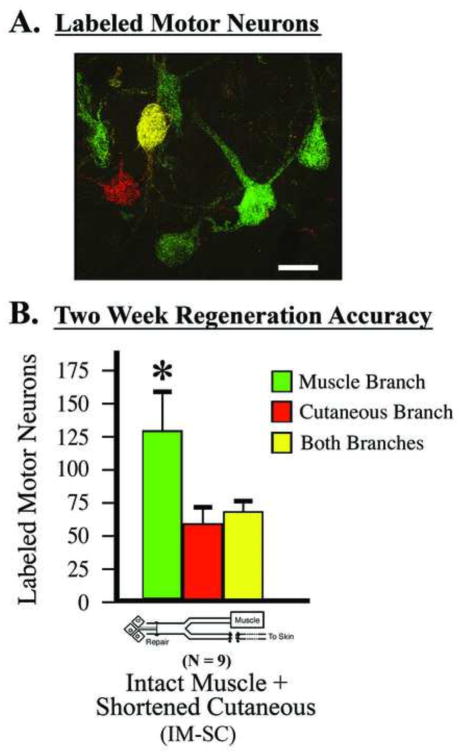
Figure 1.
Surgical model used to quantify motor neuron regeneration accuracy. A) Retrogradely labeled motor neurons in the Intact-muscle:shortened-cutaneous (IM-SC) surgical model. Single labeled motor neurons are quantified as projecting solely to either the terminal muscle branch or cutaneous branch (i.e., either green or red), while the double-labeled motor neurons are quantified as projecting to both branches (i.e., green and red, appearing as yellow). Size bar = 25 μm. B) Quantification in this model system two weeks after parent femoral nerve repair shows a significant preference for motor neurons to project to the terminal muscle branch compared to the cutaneous branch; mean ± SEM, paired t-test (t=2.67, p=.02).

