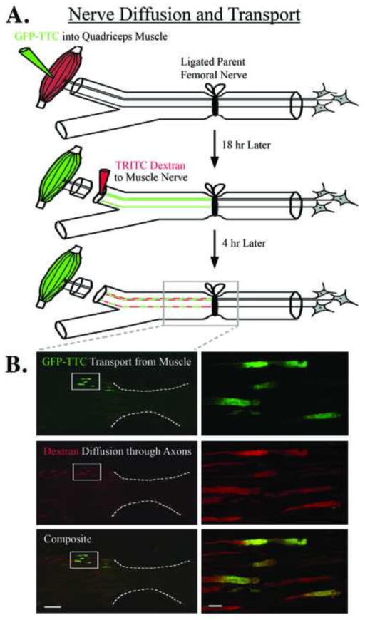
Figure 2.
Muscle-derived signals accumulate at a proximal nerve repair site. A) GFP-labeled tetanus toxin c-fragment (GFP-TTC) was injected into quadriceps muscle during the same surgery that ligated the parent femoral nerve. Eighteen hours later TRITC-labeled dextran was applied to the distal terminal muscle branch, and 4 hours after that the nerve was processed for histology. B) Upper panels show accumulation of the green GFP-labeled tetanus toxin c-fragment at the ligation site indicating that retrograde transport has continued in the denervated nerve over this time period; white box shown at higher magnification on the right for all panels, white dotted line indicates ligation site of the parent femoral nerve. Middle panels show that TRITC-labeled dextrans, which travel via diffusion driven movement, also accumulate at the repair site and are confined to specific axonal compartments and subsequent bands of Bungner which indicate that these compartments act as diffusional barriers. The lower panels are an overlay showing both the TRITC-dextran and the GFP-TTC labels. Because the TRITC-dextran was applied to the entire muscle branch it would be expected to label most of the axons within that branch, whereas the GFP-TTC would be expected to be restricted to fewer axons and only represent those motor neuron axons that were exposed to GFP-TTC due to the muscle injection. Size bar on the left = 100 μm, and on the right = 10 μm.

