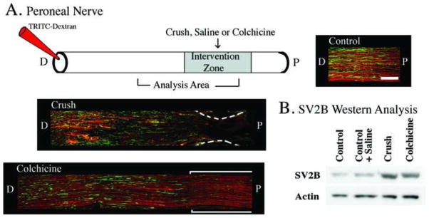
Figure 3.

Methods to disrupt diffusion driven movement and/or active axonal transport. These experiments utilized histological and Western blot analysis using SV2B (synaptic vesicle protein 2B) as an example of a protein that moves via active axonal transport, and labeled dextrans as an example of a diffusion driven molecule. A) The upper panel depicts the application of saline (control), nerve crush, or colchicine to the peroneal nerve. Eighteen hours later TRITC-dextrans were applied distal to that intervention zone to assess diffusion driven movement, and four hours later the nerve was harvested for histological and Western analysis. Longitudinal histological sections of peroneal nerve are shown and illustrate that both TRITC-dextran and SV2B are present throughout the nerve after saline application (Control, right upper panel). However, when a nerve crush was applied (middle panel) both TRITC-dextran and SV2B stopped abruptly at the crush zone, indicating disruption of both diffusion driven movement and active axonal transport. When colchicine was applied to the nerve (lower left panel) SV2B transport was disrupted at the application zone but diffusion driven movement of the TRITC-dextran was not affected. Size bar in upper right panel (Control) represents 200 μm and applies to all panels. B) Western analysis verifying the immunohistochemistry data that both a nerve crush or the application of colchicine resulted in the accumulation of SV2B at the intervention zone. N=2 animals in each group; these experiments were replicated a minimum of three times using different animals (i.e. biological replicates).
