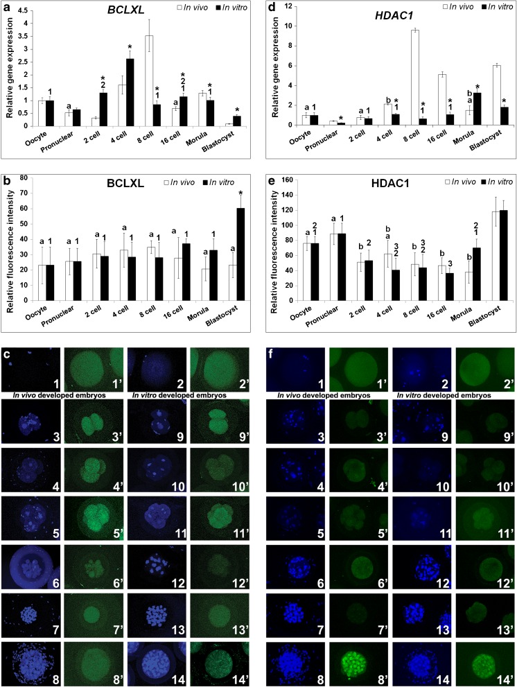
Fig. 1.
Relative expression levels of BCLXL and HDAC1 in the in vivo and in vitro developed rabbit embryos. a & d Relative mRNA expression. b & e Relative protein expression. c & f Protein localization by immunocytochemistry. In c & f (1 & 1′) oocyte, (2 & 2′) pronuclear embryo, (3 & 3′ and 9 & 9′) 2-cell embryo, (4 & 4′ and 10 &10′) 4-cell embryo, (5 & 5′ and 11 & 11′) 8-cell embryo, (6 & 6′ and 12 & 12′) 16-cell embryo, (7 & 7′ and 13 &13′) Morula and (8 & 8′ and 14 &14′) Blastocyst. Figures from 3 to 8 are in vivo developed embryos and figures from 9 to 14 are in vitro developed embryos. DAPI (1–14) and FITC (1′–14′) emission signals for respective developmental stages are shown side by side. Asterisk (*) indicates significant difference between in vitro and in vivo developed embryos in respective developmental stages (P < 0.05). Identical superscripts (alphabets for in vivo developed embryos and numerals for in vitro developed embryos) indicate similar levels of expression between developmental stages (P < 0.05)

