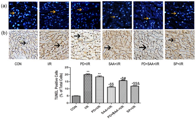
Figure 3. Effects of SAA on apoptosis of I/R myocardium in vitro.
A representative photomicrograph of DAPI-stained (Figure 3a) and TUNEL (Figure 3b) cardiomyocytes were showed. After 2 h reperfusion, the heart tissure were sectioned for analysis of anti-apoptotic effect of SAA, PD and SP, cardiomyocytes were stained with DAPI, and the ratio of TUNEL-positive cardiomyocytes was calculated. *P<0.05, **P<0.01 versus CON group, $P<0.05, $$P<0.01 versus I/R, #P<0.05, ##P<0.01 versus SAA+I/R, &P<0.05, &&P<0.01 versus PD+SAA+I/R. All data were expressed as mean ±SEM, n = 6. All data were expressed as mean ±SEM, n = 6. Cells were examined by light microscopy (200×magnification). Yellow allows indicate DAPI-stained nucleus, black allows indicate TUNELpositive caryons.

