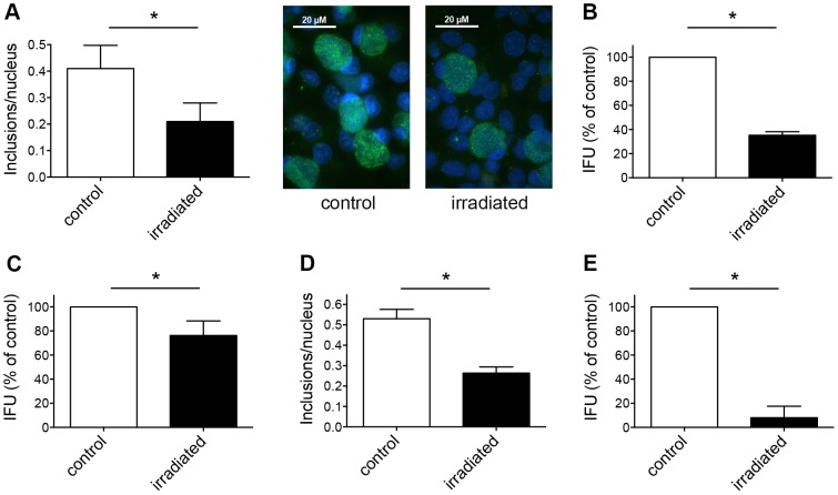
Figure 2. Irradiation of chlamydial EBs reduces their infectivity on host cells.
(A) C. trachomatis EBs (MOI 1) were either irradiated or not prior to infection of HeLa monolayers. Cultures were incubated for 43 hours, fixed, and immunolabelled with anti-chlamydial LPS (green) and DAPI (blue). Frequency of inclusions per nucleus was calculated (mean ± SD; * p<0.05; n = 3; t test). Representative microscopic pictures at 1000× magnification are shown additionally in the panel. (B) Cultures were collected and subjected to sub-passage titer analysis. Inclusion forming units (IFU) are shown as percent of control (mean ± SD; * p<0.0001; n = 3; t test). (C) C. pecorum EBs were irradiated prior to infection of HeLa monolayers. Non-irradiated EBs were used as controls. Cultures were collected and subjected to sub-passage titer analysis. Inclusion forming units (IFU) are presented as percent of control (mean ± SD; * p<0.05; n = 3; t test). (D) C. pecorum EBs were irradiated or not prior to infection of Vero monolayers. Cultures were incubated for 43 hours, fixed, and immunolabelled as shown in panel A. Frequency of inclusions per nucleus was calculated (mean ± SD; * p<0.0025; n = 3; t test). (E) Cultures from panel D were collected and subjected to sub-passage titer analysis. Inclusion forming units (IFU) are shown as percent of control (mean ± SD; * p<0.0001; n = 3; t test).

