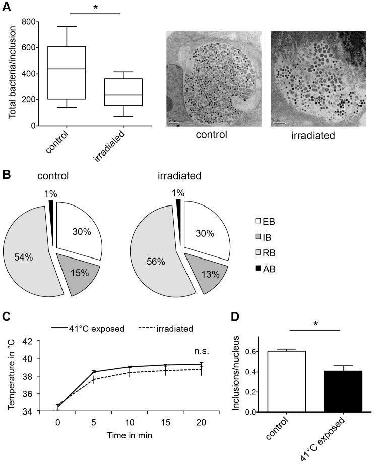
Figure 5. Thermal effects of irradiation are responsible for the reduced chlamydial infectivity.
(A) C. pecorum-infected Vero cells were irradiated 40 hpi. Non-irradiated, C. pecorum-infected cells are controls. Cultures were fixed 43 hpi with glutaraldehyde, and further processed as described in Material and Methods in the transmission electron microscopy section. The total number of bacteria in ten inclusions per condition was counted: controls (in sum 4318 bacteria) and irradiated inclusions (2524 bacteria). Shown are the means ± SD of ten chlamydial inclusions (* p<0.05, n = 10; t test). The mid-line shows the median, the box represents the 25th to 75th percentiles whereas the whiskers show the minimum to maximum values. Representative inclusions are shown additionally in the panel. (B) Chlamydial bacteria within ten inclusions (from A) were split according to their morphology into elementary bodies (EB), intermediate bodies (IB), reticulate bodies (RB) and aberrant bodies (AB). The graphs show the distribution of each maturation stage per condition. (C) 1 mL of cell culture medium in a well of a 24-well plate was irradiated or the resulting temperature enhancement due to irradiation was mimicked using a water bath at 41°C (41°C exposed). Shown is the intra-well temperature at the indicated time points (mean ± SD; n.s. non significant, n = 3). (D) Vero cells were infected with C. pecorum, incubated for 40 h and placed in a water bath at 41°C for 20 min (41°C exposed). In parallel, C. pecorum-infected Vero cultures were placed in a water bath at 37°C for 20 min and were used as controls. Cultures were subsequently fixed at 43 hpi and immunolabelled with anti-chlamydial LPS (green) and DAPI (blue). Frequency of inclusions per nucleus was calculated (mean ± SD; * p<0.005; n = 3; t test).

