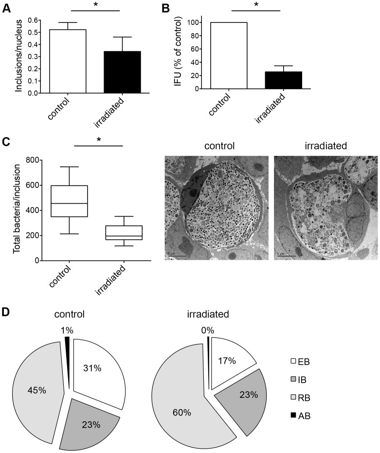
Figure 6. Triple dose of irradiation enhances the effect on chlamydial inclusions and their maturation stages.
(A) C. trachomatis-infected HeLa cells were irradiated three times at 24, 36 and 40 hpi for each 20 min. Non-irradiated, C. trachomatis-infected cells were used as controls. Cultures were fixed 43 hpi and immunolabelled with anti-chlamydial LPS (green) and DAPI (blue). Frequency of inclusions per nucleus was calculated (mean ± SD; * p<0.01; n = 3; t test). (B) Infected HeLa cultures were collected and subjected to sub-passage titer analysis. Inclusion forming units (IFU) are presented as percent of control (mean ± SD; * p = 0.0001; n = 3; t test). (C) Cultures were fixed 43 hpi and further processed to transmission electron microscopy. The total number of bacteria in ten inclusions per condition was counted: controls (in sum 4627 bacteria) and irradiated inclusions (2179 bacteria). Shown are the means ± SD of chlamydial inclusions (* p<0.0005, n = 10; t test). The mid-line shows the median, the box represents the 25th to 75th percentiles whereas the whiskers show the minimum to maximum values. Representative inclusions are shown additionally in the panel. (D) Chlamydial bacteria within ten inclusions (from C) were split according to their morphology into elementary bodies (EB), intermediate bodies (IB) and reticulate bodies (RB). The graphs show the distribution of each maturation stage per condition.

