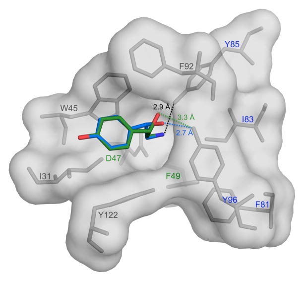
Fig. 6.
Octopamine modeled in the Bla g 4 binding site. S-octopamine is shown in green, while R-octopamine is shown in blue. Hydrogen bonding distances from O7 of S-octopamine and R-octopamine to the hydroxyl group of Y96 is 3.3 Å and 2.7 Å respectively. The distance from N8 of S/R-octopamine to the hydroxyl group of Y85 is 2.9 Å. Residues labeled in blue are conserved between Bla g 4 and Per a 4, while residues labeled in green are similar.

