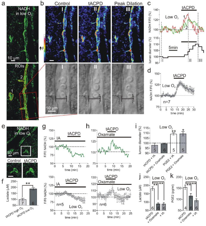
Fig. 3.

Glycolysis and lactate release is critical for astrocyte-mediated dilations. (a) Arteriole and astrocyte NADH image in low O2; bottom shows ROIs for astrocyte compartments (1 and 2) and vessel (box). (b) Pseudo color NADH (top) and diameter (bottom) changes caused by t-ACPD at time points I-III in c. Vertical dotted line (II and III) indicates previous position of vessel wall. (c) Astrocyte NADH (top) and lumen diameter (bottom) in response to t-ACPD; same experiment as a and b. (d) Summary of NADH increase from mGluR activation in low O2. (e) Top: NADH image of an arteriole and astrocyte. Lower: soma close-up showing t-ACPD causes a diffuse increase in NADH. (f) t-ACPD increases extracellular lactate most in low O2. (g) t-ACPD decreases astrocyte NADH during glycolysis inhibition; top: single experiment; bottom: summary. (h) mGluR activation increases astrocyte NADH during LDH inhibition; top: single experiment; bottom: summary. (i) Summary showing t-ACPD fails to dilate vessels in oxamate or IA and PGE2 rescues vasodilation in these compounds. (j and k) The increase in lactate (j) and PGE2 (k) caused by t-ACPD is significantly less in oxamate and IA.
