
Fig. 4.
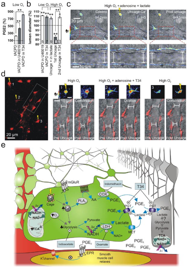
Raising PGE2 levels by inhibiting PGT changes the polarity of astrocyte-mediating vasomotion. (a) PGE2 levels were further elevated by mGluR activation in low O2 when PGT was inhibited by U46619 or T34. (b) Summary data of vasomotion during PGT manipulation. (c) Uncaging Ca2+ in high O2 causes vasodilation in exogenous lactate. Top panels show the astrocyte Ca2+ signal change (pseudo colour) from uncaging. Lower panels show close up of vessel lumen. (d) In high O2, astrocyte-mediated vasoconstriction is converted to vasodilation during PGT blockade. Left: astrocytes and endfeet circumscribing an arteriole. Ca2+ is uncaged in 3 astrocytes separated in time; box indicates the vessel region examined on the right. Right: vasomotions corresponding to the separate uncaging events. Small pseudo colour images show the Ca2+ signal change from uncaging in each astrocyte. Lower images of the vessel and endfeet (red) show that the vasomotion switches polarity when PGTs are blocked. (e) Diagram of the supported model.