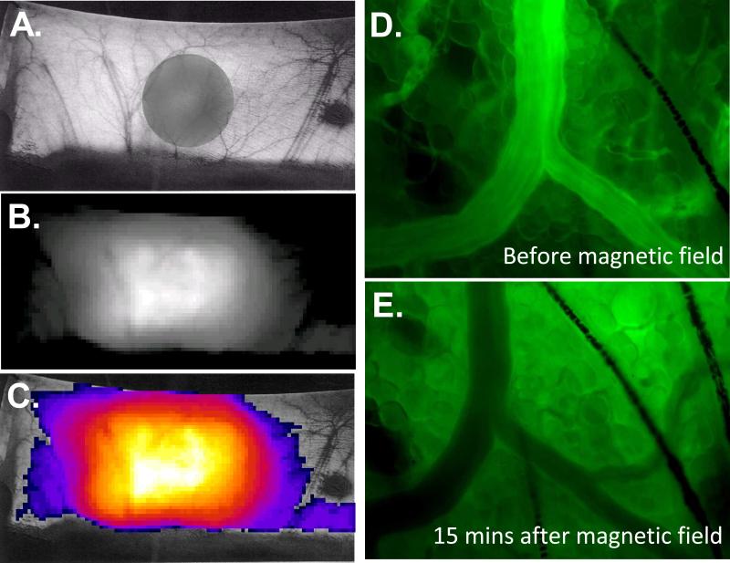
Figure 7.
A. Hamster dorsal depilated skin, gray shadow projected by the circular magnet placed underneath the lifted skin. B. Intensity collected from rhodamine labeled L35-PMNPs one hour after injection using a 500-720 nm emission filter with exposure time of 1 sec. C. Pseudo-color accumulation of rhodamine labeled L35-PMNPs. D. Intravascular fluorescent image after infusion of rhodamine labeled L35-PMNPs before application of magnetic field. E. Intravascular fluorescent image after 15 min of application of magnetic field. Microscopic fluorescence images were taken to identify the presence of rhodamine labeled L35-PMNPs in the tissues.

