Fig. 1.
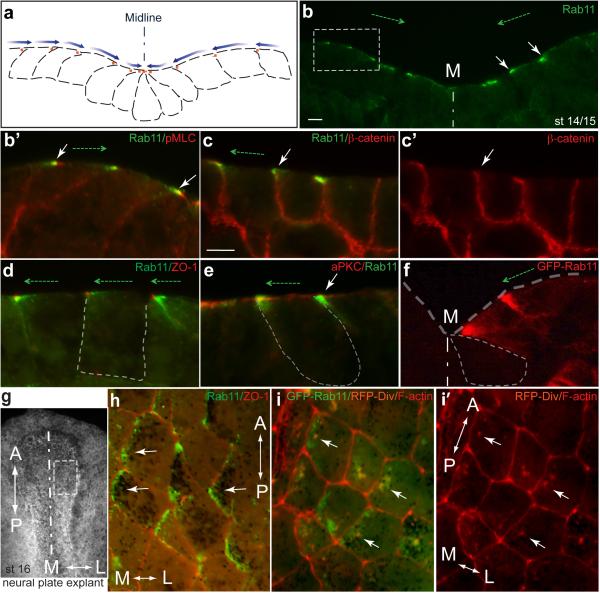
Rab11 distribution reveals planar polarity along the mediolateral axis of the Xenopus neural plate. a, b, Scheme (a) and a representative transverse cryosection (b) of the neural plate stained with anti-Rab11 monoclonal antibodies at stage 14/15. b’, higher magnification is shown for image within the white square in b. Costaining for phospho-myosin II light chain (pMLC, b’) reveals cell boundaries. c-e, Embryo sections were co-stained with Rab11 and β-catenin (c-c’), ZO-1 (d) and aPKC (e) antibodies at higher magnification. f, Polarized localization of exogenous Rab11. Four-cell embryos were injected with 100 pg of GFP-Rab11 RNA and stained with anti-GFP antibodies at midneurula stages (st. 16). b-f, White arrows point to polarized Rab11 distribution. Dashed green arrows, and blue arrows in (a), directed towards the midline (M) indicate cell and tissue polarity. Neural plate and individual cell boundaries are shown by broken lines. g-i, En face view of the neural plate with polarized Rab11 and Diversin (white arrows). g, dorsal view of a neural plate explant (stage 16) with white rectangle indicating approximate location of images in h, i. A, anterior, P, posterior, M, medial, L, lateral. h, Endogenous Rab11 is medially enriched along the ML axis. i, i’, Both exogenous GFP-Rab11 (green vesicles) and Diversin-RFP (i’, cytoplasmic red staining) show medial polarization along the ML axis (white arrows) and partly colocalize. Antibody specificity is indicated at the upper right corner of each panel. The AP axis is indicated. Scale bar in b and c (also refers to d-f) is 10 μm.

