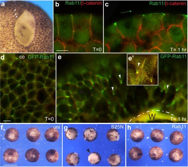
Fig. 6.
Planar polarity of Rab11 endosomes during epithelial wound healing. a, Representative blastula embryo (stage 9), 30 min after wounding. Arrowheads point to polarized cells. b, Rab11 staining in control ectoderm, c, Representative ectodermal explant with Rab11 polarization relative to the wound position. Costaining with α-catenin antibodies indicates cell boundaries. Dashed green arrow indicates the direction toward the wound. d, e, Live images of GFP-Rab11 polarization (white arrows) in ectoderm during wound healing. d, control ectoderm; e, explant with wound healing for 1 hr. Wound (W) position is indicated by broken line. e’, Inset shows two individual cells with polarized GFP-Rab11. f-h, Rab11 is essential for wound healing in ectodermal explants. Ectodermal explants were prepared at late blastula stages from the uninjected embryos (f), or embryos injected with wild-type Rab11 (h) or Rab11S25N (g) RNAs (1 ng each), and allowed to heal for 1 hr at room temperature. Scale bars are 20 μm in (b) and (d), also refer to (c, e), and 300 μm in (f), also refers to (g, h). Results are representative of 3-5 independent experiments.

