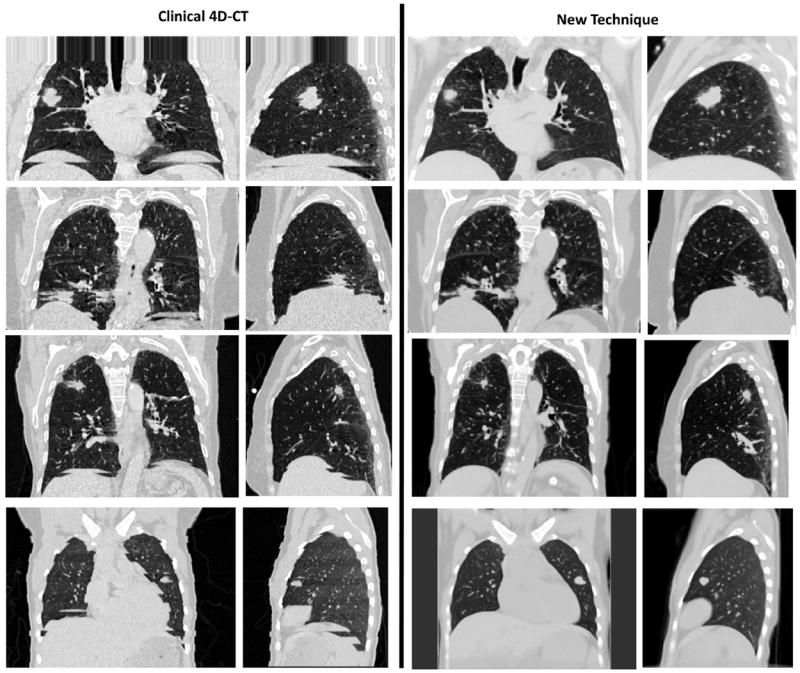
Fig. 4.

(Left column) Examples of clinical 4D-CT images are shown. (Right column) Sorting artifact–free image was generated by using the new 4D-CT method. Images at the 25th percentile tidal volume, central coronal slice, and right lung sagittal slice are shown in a lung window.
