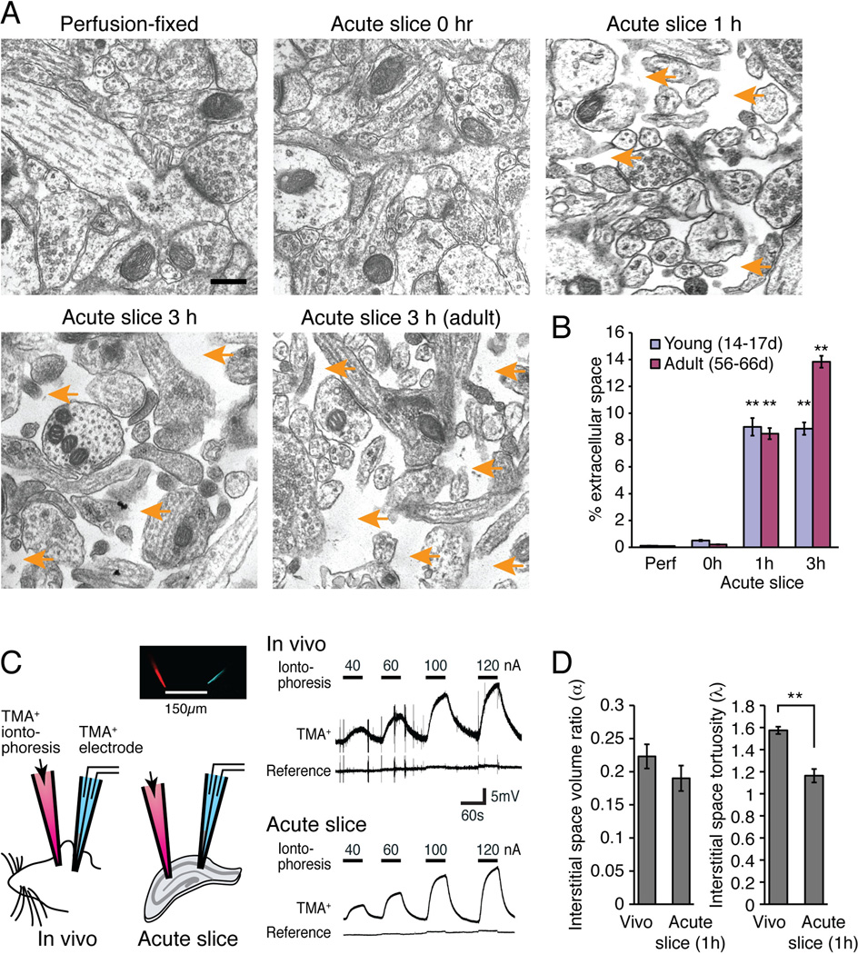
Fig. 6. Changes in the interstitial space volume and tortuosity in acute hippocampal slices.
(A) Regular EM showed that the interstitial space (orange arrows) expanded during incubation of hippocampal slices in an oxygenated aCSF. Scale bar, 0.3 µm. (B) Comparison of the relative extracellular space in perfusion-fixed (Perf) versus immersion-fixed slices at 0, 1, and 3 h after vibratome cutting taken from young (14–17-day-old postnatal pups) and adult (56–66-day-old) animals (N=40 fields from 9–18 animals, Mean ± sem, **, p<0.01, Kruskal-Wallis test with Dunn’s test against perfusion fix). (C) Real-time ionophoretic TMA+ measurement of interstitial space in slices (1 h after preparation) and in vivo using a double-barrel electrode comprised of TMA+ sensitive and K+ reference electrodes placed at 150 µm away from the TMA+ iontophoresis. (D) Histograms showing the interstitial space volume fraction (α) and tortuosity (λ) of the extracellular space (N=7–12, Mean ± sem, **, p < 0.01, t tests).

