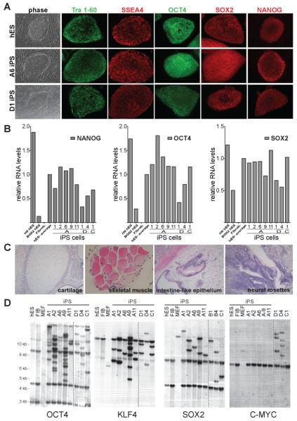
Figure 1. Characterization of DOX-inducible iPS cells derived from human fibroblasts.
A. Phase contrast picture and immunofluorescence staining of human ES (hES) cells and iPS cell lines A6 and D1 for pluripotency markers SSEA4, Tra 1-60, OCT4, SOX2 and NANOG.
B. Quantitative RT-PCR for the reactivation of the endogenous pluripotency related genes NANOG, OCT4 and SOX2 in independent iPS cell lines, hES cells and primary fibroblasts. Relative expression levels were normalized to the average expression of the two control hES cell lines.
C. Hematoxilin and eosin staining of a teratoma sections generated from A1 iPS cells.
D. Southern blot analysis of parental GM01660 fibroblasts, hES cells and iPS cells for proviral integrations of xbaI digested genomic DNA using 32P-labeld DNA probes against OCT4, KLF4, SOX2 and C-MYC.

