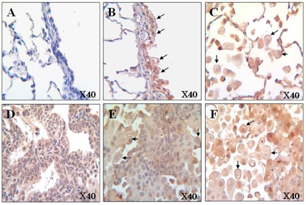
Fig. 3.

Representative image of α7 nAChR immunohistochemistry in the lung of ferrets that were non-exposed (Panel A) and exposed to NNK for 26 weeks [bronchiolar epithelial cells (Panel B), alveolar macrophage (Panel C), adenocarcinoma (Panel D), squamous cell carcinoma (Panel E), and tumor associated macrophage (Panel F)] at 40X magnifications. The arrows indicated the expression of α7 nAChR (strained in reddish-purple) on the cell membranes.
