
Figure 1.
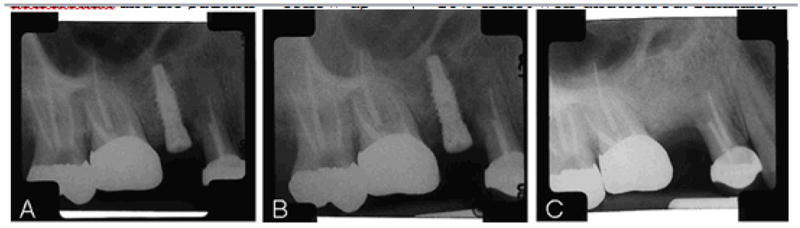
(A) Maxillary right second premolar immediately after implant placement. (B) Failing implant, maxillary right second premolar. Notice the radiolucent area around the metal. (C) Healed implant site 8 months after implant removal and 4 months after teriparatide infusions were started.