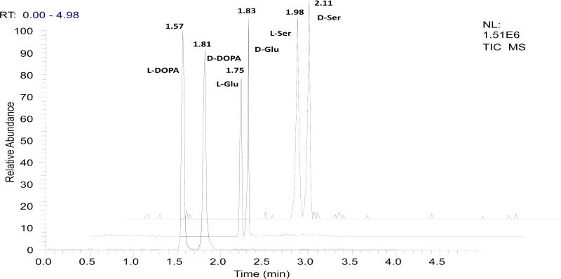
Fig 3.
TIC electropherograms from the proposed chiral MCE-MS separation of neuroactive compounds, i.e. DOPA, glutamic acid (Glu), and serine (Ser). [test compound] = 100 μM (each enantiomer). Chiral MCE conditions: MCE separation channel, 4 cm long × 60 μm wide × 20 μm deep; electrokinetic sample injection, 15 s at 600V; 35nL chiral selector solution infused by a syringe pump; separation voltage, 3850V; electrospray voltage,1500V; MCE running buffer, 15mM ammonium acetate/ acetic acid buffer (pH 5.5) /methanol (1:1); chiral selector solution, the MCE running buffer containing 15 mM sulfated β-CD; MUF, the MCE running buffer at a flow rate of 100 nL /min.

