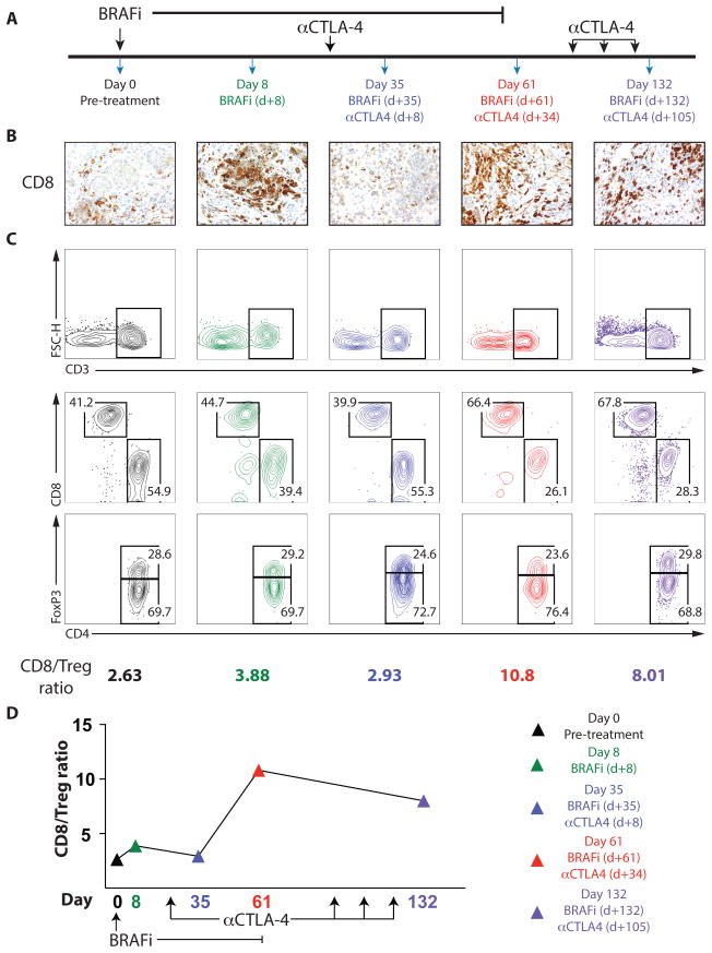
Figure 1. Combined BRAF inhibitor and anti-CTLA-4 administration leads to prolonged antitumor immunity in a patient with metastatic melanoma.
A patient with metastatic melanoma was treated with combined BRAF-targeted therapy plus CTLA-4 blockade. A, Timeline showing treatment schedule and biopsies B, CD8+ T-cell infiltrate was determined by IHC (40x magnification). C and D, Immune cells isolated from tumors were analyzed by flow cytometry. (C, Top) CD3+ lymphocytes. FSC-H, forward scatter height. (C, Middle) Populations of CD4+ and CD8+ lymphocytes pregated on CD3+ cells. (C, Bottom) Percentage of CD4+FoxP3+ T regulatory cells (Treg) and CD4+FoxP3− non-Tregs, pregated on CD3+CD4+ T cells. The ratio of CD8+ T cells to CD4+FoxP3+ Treg cells is shown (C) and plotted versus time (D).

