
Figure 1. Pulmonary vascular changes in PAH includes infiltrating adaptive and innate immune cells.
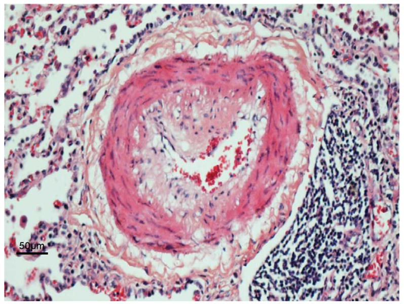
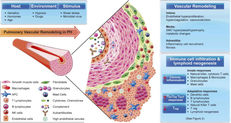
In the top panel is a representative histophathology of a vessel with severe neointimal formation represented below by a diagrammatic illustration. The histopathology shows a single endothelial layer and an eccentric neointima (pale pink) that contains cells that have markers of inflammatory cells and others that stain with markers of smooth muscle but appear poorly differentiated. The medial muscular layer is expanded and there is an abundant adventitial layer. This vessel is decorated with complement and autoantibodies, infiltrated by neutrophils in the lumen attacking the vessel wall and other inflammatory cells binding to the endothelium and infiltrating. The neointima is comprised of pale cells and matrix and infiltrating T and B cells and in the adventitia there are dendritic cells, macrophages and mast cells and in the periadventitial space tertiary lymphoid follicles characterized by T cells, B cells and plamacytoid dendritic cells (APC).