
Fig. 2. HIV-infected leukocyte interaction with epithelial cells: attachment and directional viral shedding.
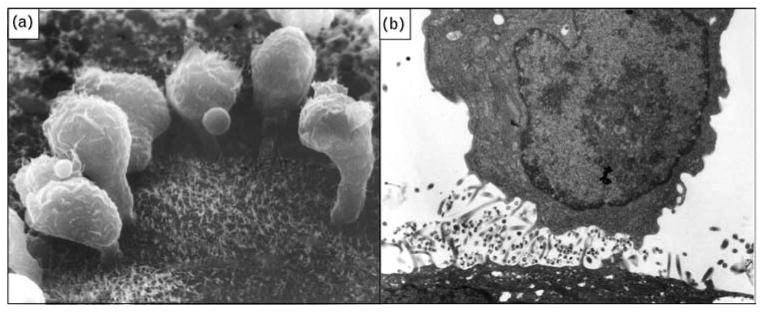
(a) Scanning electron micrograph showing HIV-infected lymphocytes adhering to the surface of an epithelial cell (magnification × 10000). (b) Transmission electron micrograph of HIV-infected macrophage from semen releasing virus after contact with a genital tract epithelial cell (magnification × 15 000). Original photographs provided by David M. Phillips with permission from the Population Council, New York. Part (a) reproduced from [4].