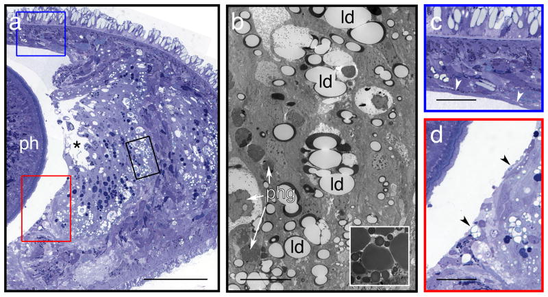
Figure 3. Artefacts not caused by fixation.
(a) Light micrograph of 0.5 μm cross-section (ph: pharynx) through an asexual worm. Dehydration of specimens by the “rapid, cold” schedule described in the Experimental design section results in undesirable extraction of lipids from specimens. This effect is most noticeable in the lipid droplets that are abundant in the digestive tract of the worm, giving it a frothy appearance. (In the comparative experiments described in Figure 1, all specimens dehydrated by the rapid cold method exhibited such features). The large space indicated by an asterisk is the gut lumen, the wall of which was ruptured by mechanical damage, rather than fixation or dehydration – see c and d, which illustrate the blue- and red-boxed regions, respectively. (b) Transmission electron micrograph of a 70 nm section, adjacent to area enclosed by black box in (a). At this magnification, a thin rim of unextracted lipids, stained dark by OsO4, can be seen. Inset shows lipid droplets in a similar region of the digestive tract, from a specimen dehydrated by the gradual, room-temperature method described in the main protocol. All specimens dehydrated gradually at room temperature exhibited such full droplets – see Figures 1a-c. Arrows indicate some of the many phagosomes present (phg). The apparent shrinkage or condensation of electron-dense materials evident in larger phagosomes was found in similar structures in all worms, and is not due to the dehydration process used here. (c, d) Mechanical damage: partial tearing of the surface layer of the pharyngeal cavity. A thin layer of epithelial tissue normally covers the lumenal surface of the body wall around this cavity. In this specimen, such tissue is present over the dorsal surface of the cavity (panel c, white arrowheads), but missing from the lateral surface (panel d, black arrowheads). Some tissues underlying the surface layer also appear to have been torn away, such as part of the gut epithelium (panel a, *). This suggests mechanical damage to the specimen, likely during primary fixation at the cutting step (Step 5). Scale bars – a: 100 μm; b (full panel and inset): 10 μm; c, d: 25 μm.

