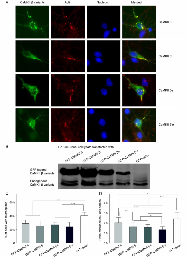
Figure 2.

Colocalization of CaMKIIβ variants with F-actin in microspikes. A. CaMKIIβ variants preferentially localized to F-actin-rich structures: Confocal images of E-18 cortical neurons at 4 DIV with mGFP tagged CaMKIIβ variants (green) and F-actin labeled with fluorescent conjugated phalloidin (red). Colocalization can be seen as yellow in the merged images. B. Enrichment of GFP-tagged proteins as indicated in microspikes (ratio of microspike/soma mean intensity) *P < 0.05; ***P < 0.001; compared with GFP-actin. ##P < 0.01; ###P < 0.001; compared with CaMKIIβ. @P < 0.05; compared with CaMKIIβ’. %P < 0.05; compared with CaMKIIβe. C. Microspikes-containing cells percentage of respectively transfected E-18 cortical neurons. **P < 0.01; ***P < 0.001; compared with GFP-actin. D. Western blot analysis of E-18 cortical neuronal culture lysates 5 DIV after mGFP tagged CaMKIIβ variants transfection (upper panel). Endogenous CaMKIIβ were identified in cultured E-18 cortical neurons (lower panel).
