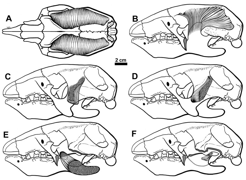
Figure 10.
Reconstructions of the M. temporalis in dorsal view ( A) and lateral view ( B), the M. masseter profunda ( C), M. zygomaticomandibularis ( D), M. pterygoideus medius ( E) and M. pterygoideus lateralis ( F). In B the mandibular coronoid process is represented as if the muscle inserting on it was transparent. In C and D, the zygomatic arch and cranial ligaments are shown as if transparent. In E, both the anterior and posterior portions of the zygomatic arch are cut (indicated by shading), as well as the ascending portion of the coronoid process. The mandible is depicted as transparent to show the insertion of the muscle on the medial side. In F the portions of the zygomatic arch are cut as in E, and the shape of the pterygoids flanges are shaded grey. The mandible is also depicted as transparent, with the insertion of the M. pterygoideus lateralis shows attaching to the medial side of the condylar process. This muscle has been restored with two heads as is the case in tree sloths.

