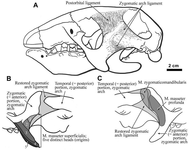
Figure 3.
The cranium of Hapalops in lateral view showing the restored cranial ligaments ( A). Enlargements of the lateral ( B) and medial ( C) surfaces of both the anterior and posterior portions of the zygomatic arch in Hapalops are also shown, along with the zygomatic arch ligament, and the locations of the scars of muscle and muscle head origins that are indicated by differentially shaded areas.

