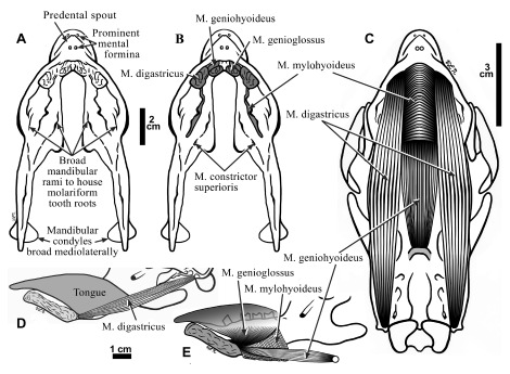
Figure 7.

Ventral views of the mandible of Hapalops showing suprahyoid muscle scars ( A) and with areas of muscle origins shaded ( B); 2 cm scale. These muscles are reconstructed in ventral view ( C) and lateral view ( D, E). In D and E, the tongue position has been estimated to show it relative to that of the oral musculature; 1 cm scale.
