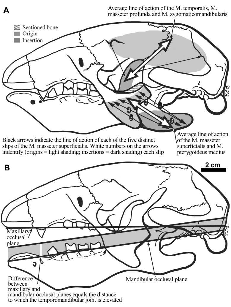
Figure 8.
The cranium of Hapalops is shown in lateral view ( B), illustrating the differential height of the craniomandibular joint for the skull and mandible. In A, the lines of action of the five individual parts, and a composite line of action of the M. masseter superficialis, and the anterior and posterior as well as a composite line of action for the M. temporalis are illustrated.

