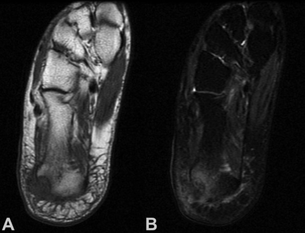
Figure 1. A 40-year-old woman with pain at the right calcaneus. Coronal (A) T1-weighted MR image showing a isointense to muscle lesion. involving the calcaneus with soft tissue mass; (B) T2-weighted MR imaging showing heterogeneity of the lesion with foci of hypointensity. Biopsy showed clear cell sarcoma. Wide resection with partial calcanectomy and local flap wound coverage was done, without evidence of local recurrence or distant metastases 3 years after diagnosis and treatment.

