Figure 1.
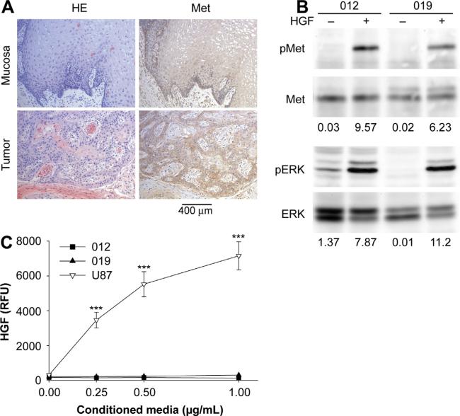
Met signaling in human oral cancer. (A) H&E staining and Met immunohistochemistry revealed that human oral tumor cells express higher levels of Met compared to adjacent normal mucosa (n = 5). (B) Lysates from JHU-SCC-012 or JHU-SCC-019 cells treated without (−) or with (+) 100 ng/mL HGF for 5 min were examined by western analysis for Met, ERK and phosphorylation of Met (pMet, Y1234/1235) and ERK (pERK). Representative results from triplicate experiments are shown. The relative density of pMet/Met and pERK/ERK is indicated beneath the respective blots. (C) HGF-ELISA detected no HGF in conditioned media from JHU-SCC-012 and JHU-SCC-019 cells whereas U87 cells were positive for HGF secretion. Values are expressed as the average mode ± SEM (*** p < 0.001; ANOVA) from triplicate experiments.

