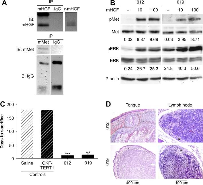
Figure 2.
Human HNSCCs form orthotopic tumors in mice. (A) Western analysis of immunoprecipitations from mouse tongue lysates using antibodies for mouse HGF (mHGF), mouse Met (mMet) or goat IgG detected mHGF (top panel) and mMet (lower panel) is this tissue. Recombinant mouse HGF (r-mHGF) was used as positive control. Representative results from triplicate experiments are shown. (B) Sera starved JHU-SCC-012 and JHU-SCC-019 cells were treated without (−) or with 10 ng/mL or 100 ng/mL HGF and examined by western analysis for pMet, Met pERK, ERK and β-actin. Representative results from triplicate experiments are shown. The relative density of pMet/Met of pERK/ERK compared to β-actin is shown beneath the respective blots. (C) Mean survival of mice (n = 5) injected orthotopically into the lateral tongue with saline, immortalized oral keratinocytes (OKF-TERT1), JHU-SCC-012 or JHU-SCC-019 cells (1 × 10⁶). Values are expressed as the average mode ± SEM (*** p < 0.001; ANOVA) of triplicate experiments. The graph represents days to sacrifice at the end of protocol (6 months) or following 20% weight loss. (D) Histological analysis of tumor bearing mice by H&E staining of the tongue and lymph node. Asterisks denotes tumor in lymph node.

