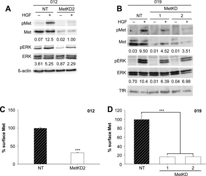
Figure 4.
Met knockdown in JHU-SCC-012 and JHU-SCC-019 cells. JHU-SCC-012 (A) or JHU-SCC-019 (B) cells infected with recombinant lentivirus expressing Met knockdown shRNA (1 or 2) or a non-targeting (NT) shRNA were treated without (−) or with (+) HGF and examined by Western analysis for pMet (Y1234/1235), Met, pERK, ERK, β-actin or Transferrin Receptor (TfR) levels. Representative results from triplicate experiments are shown. The relative density of pMet/Met of pERK/ERK compared to β-actin or TfR is shown beneath the respective blots. Flow cytometry detected reduced cell surface Met levels in JHU-SCC-012 (C) and JHU-SCC-019 MetKD (D) cell lines. Values are expressed as the average mode ± SEM (*** p < 0.001; ANOVA) of three independent experiments.

