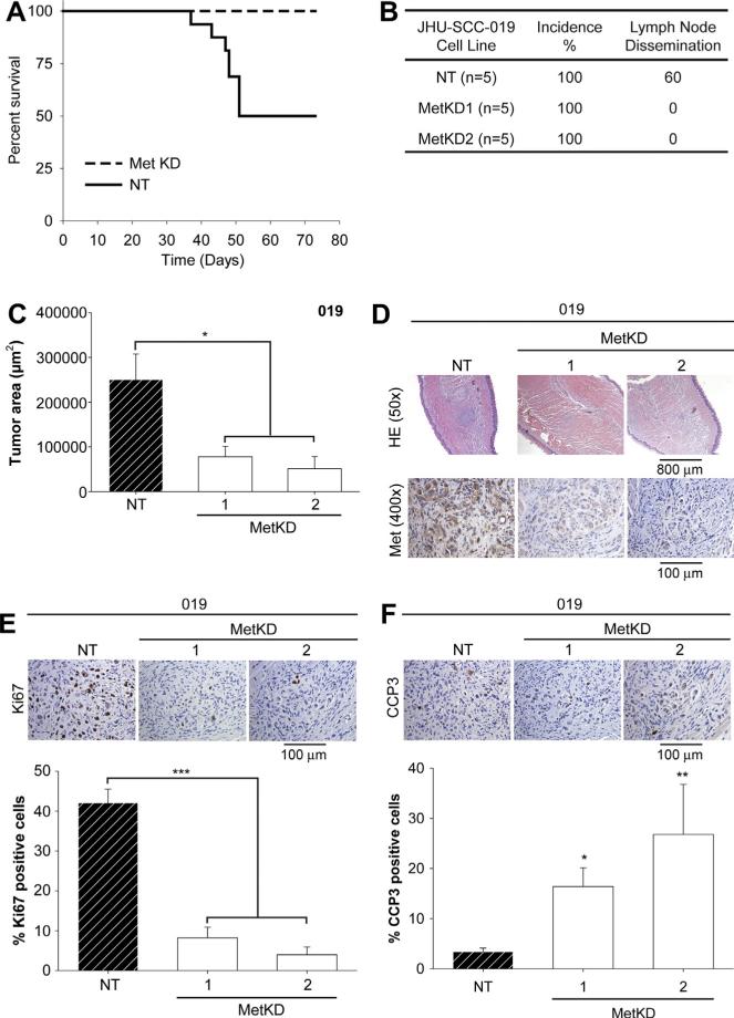
Figure 7.
Reduced tumor burden In vivo following MetKD. Mice were injected orthotopically into the lateral tongue with JHU-SCC-019 MetKD (n = 5) or control (NT) cells (n = 5). Survival rates were examined every day for up to 75 days and are plotted as a Kaplan Meier curve (A). 30 days post injection, serial sections of paraffin-embedded tongues and cervical lymph nodes were H&E stained and analyzed for tumor dissemination (B) and size (C). Adjacent slides were processed for Met (D), Ki67 (E) and cleaved caspase 3 (CCP3) (F) staining. Morphometric analysis indicated a significant decrease in tumor size and Ki67 staining with increased CCP3 per high powered field in MetKD-derived versus NT-derived tumors (* p < 0.05, ** p < 0.01, *** p < 0.001; ANOVA, n = 5). Representative images are shown.

