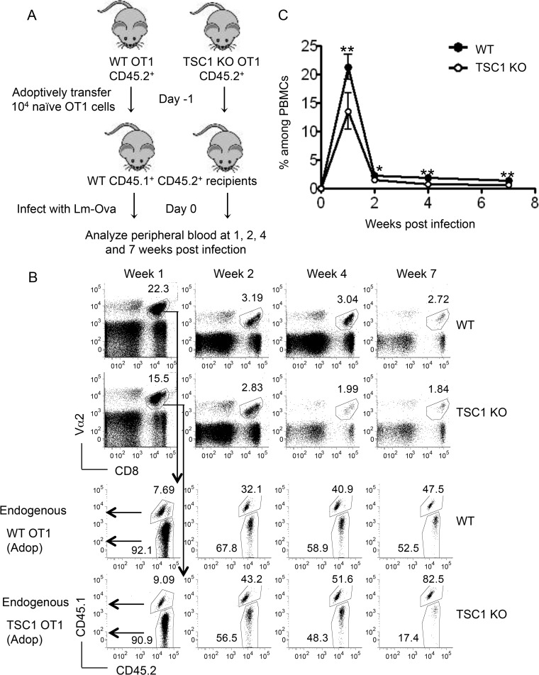
FIG 2.
TSC1 deficiency impairs antigen-specific CD8 responses. (A) Schematic representation of the experimental design showing individual adoptive transfers of naive WT OT1 or TSC1 KO OT1 cells (CD45.1− CD45.2+) into WT CD45.1+ CD45.2+ recipients. (B) Representative fluorescence-activated cell sorter analysis of peripheral blood samples showing percentages of Vα2+ CD8 cells among the total PBMCs (top). The analysis on the bottom was gated on this Vα2+ CD8+ population, and the adoptively transferred (CD45.1− CD45.2+) and endogenous (CD45.1+ CD45.2+) populations at the postinfection times indicated are shown. (C) Percentages of WT and TSC1 KO cells among the total PBMCs at the postinfection times indicated. The percentage of WT cells, for instance, was calculated as a product of the percentage of Vα2+ CD8+ cells among the total PBMCs and the percentage of WT cells (CD45.1− CD45.2+) within the Vα2+ CD8+ population. The mean ± the standard error of the mean was calculated for five mice per group. The data shown are representative of three independent experiments. *, P < 0.05; **, P < 0.01 (Student t test).

